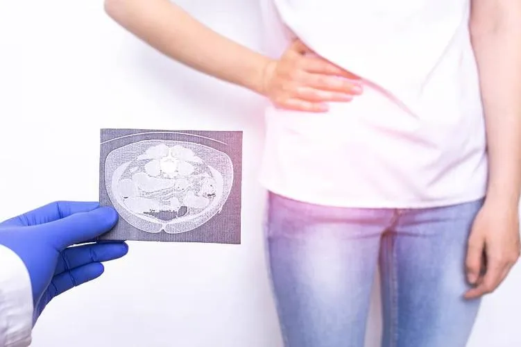
Reasons to get a routine imaging of suspected appendicitis
October 3, 2022
Acute Appendicitis (AA) is a common cause of acute abdominal pain and must be surgically operated on in complicated cases like a perforated appendix or pregnancy to prevent rupture and subsequent sepsis that could be life-threatening. For this, prompt and accurate diagnosis is crucial either to carry on with appendectomy (surgical removal of the appendix) as well as to prevent false diagnosis that often leads to negative appendectomy (removal of the appendix as a precaution or by a mistaken diagnosis of appendicitis).
Diagnosis
Appendicitis is diagnosed by the following methods:
- Clinical Diagnosis. This includes typical blood tests for CRP levels, WBC count, urine test, platelet count, and so on.
- Ultrasonography (USG)
- Radiological imaging
- CT scan
- MRI
Importance of imaging in AA diagnosis
Ø Prevention of delay in proper diagnosis that may lead to a perforated appendix
Ø Reducing the Negative Appendectomy Rate (NAR)
Ø Reducing mortality due to rapid identification and following treatment
The traditional methods of the scoring system and clinical diagnosis were found to vary greatly with gender (especially for reproductively active females), age and other medical conditions like PID (pelvic inflammatory disease), Crohn’s disease, and appendix neoplasm that have similar symptoms.
Radiology
Cross-sectional radiological imaging of the appendix has been increasingly helpful in diagnosing AA and is first in line for proper evaluation but has slowly become obsolete due to the development of more sensitive technologies.
USG
USG has proved to be a valuable tool in accurately visualising normal appendix in patients (up to 90 to 96%) with specificity as high as 85-100% and sensitivity levels up to 75 -95%. The benefits:
- Non-invasive procedure
- No need for radiation
- Quick results
- Easy to detect other causes of abdominal pain like the presence of ovarian cysts and abscesses, occurrence of ectopic pregnancy, etc. This helps in preventing NAR.
CT Scan
NAR has been found to be directly proportional to an increase in CT scans in AA detection, specifically because of its high rate of accuracy - 93-98%, sensitivity 87-100% and specificity 95-99%. It also eliminates the dependence on skilfulness of the operator as opposed to USG.
The benefits of CT scan in appendicitis:
- Perfect for detecting AA at the initial stages
- Scans can visualise the appendix more precisely than USG
- It shows the diameter of an inflamed appendix greater than 6mm diameter as a positive sign of AA. A Contrast CT scan clearly shows a thick-wall appendix due to inflammation.
- It also reveals other changes associated with appendicitis like the presence of fat stranding (attenuation of the fat layer in the peritoneum cavity), edema and enlargement of lymph nodes around the appendix, presence of air bubbles and abscesses (pus-filled blisters), phlegmon (inflamed soft tissue like viscera and omentum around appendix).
- It can also identify the presence of an appendicolith i.e formation of stones within the appendix.
- Helical CT scans have been found to be particularly helpful in identifying appendicitis with severe gynaecological disorders, where diagnosis via USG only seems unsatisfactory.
MRI scan
MRI is mostly employed in emergency cases like for children younger than 5 years and pregnant women, as it is safer than a CT scan due to lower dosage of radiation and higher accuracy. In pregnant women in their second and third trimesters, the distended uterus easily displaces the appendix making it almost impossible to detect by USG.
Conclusion
The importance of imaging in appendicitis detection is manifold even if it is expensive. It is also cost-effective and can prevent morbidity caused by delayed treatment, unnecessary operations and needless hospitalisation for observation. A misdirected surgery like a negative appendectomy is fairly risky.
You can easily request an appointment at Apollo Spectra Hospitals, Call 18605002244
A Helical CT scan is sensitive enough and one without the use of contrast.
But imaging practice has eliminated most of the missed diagnoses quite frequently.
Primarily, the use of contrast (administered via rectal or intravenous), cost, patient discomfort during tests, and development of allergy to iodine contrast are the chief disadvantages.
NOTICE BOARD
CONTACT US
CONTACT US
 Book Appointment


.svg)
.svg)
.svg)
.svg)


